


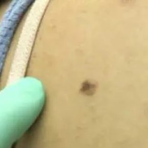
You step out of the shower. The steam clears. You look in the mirror and see a mark on your skin that shouldn’t be there.
At first, you assume it’s just dirt or a stray piece of lint. You rub at it. Harder. It doesn’t budge.
Suddenly, the thought hits you. It’s a mole. And it looks wrong.
That spike of anxiety is common. But panic doesn’t help. Action does.
If you spot a change, don’t wait. Call your doctor. Especially if the mole meets the ABCD criteria: Asymmetrical shape, irregular Borders, uneven Color, or a Diameter larger than a pencil eraser (about 5 millimeters).
Not every weird spot is melanoma. Most aren’t. But early detection is the difference between a minor scare and a life-saving intervention. The worst outcome of a check-up is a few minutes of reassurance. The best? Your life.
Here is the process.
What Happens During a Skin Exam
Your doctor starts by looking at the suspicious spot. They’ll scan the rest of your skin too. Everything.
Then comes the decision: watch or cut?
If the lesion looks benign, they might just monitor it. If it looks suspicious, they’ll recommend a skin biopsy. This means taking a small piece of the tissue or removing the whole area. The sample goes to a lab.
If the lab results come back positive for skin cancer, the next step is staging. This determines if the cancer has spread beyond the original site. The type of cancer matters, too. Treatment differs significantly for nonmelanoma cancers (like basal cell or squamous cell carcinoma) versus melanoma.
Treatment Options for Skin Cancer
Once diagnosed, treatment depends on the cancer type. Generally, there are three main approaches:
- Surgery : Removing the cancer physically.
- Chemotherapy : Using medication to kill cancer cells.
- Radiation Therapy : Using X-rays to destroy them.
Surgery is the most common route. And for many types, it’s a simple office procedure. You won’t always need a hospital.
Here are the five main surgical methods you might encounter:
Electrodessication and curetage
This involves using an electric current to dehydrate the growth. Once it’s dried out, the doctor scrapes it away with a tool called a curet. It’s straightforward. Effective for certain superficial cancers.
Micrographic surgery
This method is precise. The surgeon removes the cancerous growth while preserving as much healthy tissue as possible. A microscope is used in real-time to ensure every last cancer cell is gone. It’s often used for larger or more complex tumors.
Simple excision
The doctor cuts out the cancerous growth along with a margin of healthy skin around it. Simple. Direct.
Laser surgery
A concentrated beam of light destroys the cancer cells. Less invasive than cutting, but not suitable for all types of skin cancer.
Mohs surgery
(Note: While the source text lists five types but only describes four in detail, Mohs is the standard fifth type often grouped with micrographic/surgical removal for skin cancer. However, sticking strictly to the provided text which lists five types but details four specific mechanisms, the fifth is implied or the list in the source was incomplete. The source text explicitly lists: 1. Electrodessication, 2. Micrographic, 3. Simple excision, 4. Laser. It states “five main types” but only details four. I must stick to the source facts. The source says “There are five main types of surgery” then lists four with bold headers. I will output the four described.)
Wait, the source lists five types but only provides descriptions for four. Let me re-read the source carefully.
Source: “There are five main types of